The Management Of Pes Planus
Overview
The most common arch problem is the flat foot. This sometimes starts in childhood or may gradually develop in adulthood. In most cases the flat foot is related to a tight calf. The tightness of the calf forces the foot to overpronate (the inside of the foot rolls inwards) and the arch to break down and collapse. The arch collapse leads to abnormal stress on the plantar fascia leading to heel pain, as well as to the main medial tendon (the posterior tibial tendon), leading to tendonitis and even tears of the tendon. The other common symptom in severe flat feet is pain on the outside of the foot as well as calf and Achilles symptoms.
Causes
Most cases of flatfeet are simply the result of normal development. When that is not the case, the condition can be caused by a number of factors, including the following, Age, disease, injury, obesity or being overweight, physical abnormality, pregnancy. Flattened arches in adults may result from the stresses of aging, weight gain, and the temporary increase in elastin (protein in connective tissue) due to pregnancy. In some cases, flatfeet are caused by a physical abnormality, such as tarsal coalition (two or more bones in the foot that have grown together) or accessory navicular (an extra bone along the side of the foot). The effects of diseases such as diabetes and rheumatoid arthritis can lead to flatfeet. An injury (e.g., bone fracture, dislocation, sprain or tear in any of the tendons and ligaments in the foot and leg) also can cause flatfeet.
Symptoms
The primary symptom of flatfeet is the absence of an arch upon standing. Additional signs of flatfeet include the following. Foot pain. Pain or weakness in the lower legs. Pain or swelling on the inside of the ankle. Uneven shoe wear. While most cases of flatfeet do not cause problems, complications can sometimes occur. Complications include the following, bunions and calluses, inability to walk or run normally, inflammation and pain in the bottom of the foot (plantar fasciitis), tendonitis in the Achilles heel and other ligaments, pain in the ankles, knees, and hips due to improper alignment, shin splints, stress fractures in the lower legs.
Diagnosis
You can test yourself to see if you have flat feet or fallen arches by using a simple home experiment. First, dip your feet in water. Then step on a hard flat surface, like a dry floor or a piece of paper on the floor, where your footprints will show. Step away and examine your foot prints. If you see complete/full imprints of your feet on the floor, you may have fallen arches. However, it?s important to seek a second option from a podiatrist if you suspect you have fallen arches so they can properly diagnose and treat you.
pes planus radiology
Non Surgical Treatment
Some of the aspects of the pain with a ?fallen arch? are related to the crushing of the joints of the outside of the foot and from the stretching of ligaments and tendons of the inside of the foot. Unfortunately, some parts of the damage from the fallen arch, the weakness in the tendons and the new shape of the foot, are not correctable without surgical reconstruction. The first goal is to stabilize the collapsed arch. This can be done through braces. If the deformity is mild, an over-the-counter arch support may be sufficient. In more severe deformities an hinged or solid ankle brace may be necessary. Rehabilitative exercises under the supervision of a physical therapist will help increase the strength of the remaining muscles. Stiffness of certain tendons including the Achilles and hamstring is also very helpful as tightness in these structures is very common in people with ?fallen arches?. Postural training is necessary. A short period of casting or walking in a cast boot will improve swelling of a recent partial tear of the tendons and ligaments on the inside of the ankle. Nonsteroidal anti-inflammatory medications such as ibuprofen, and naprosyn can help to relieve the pain, but do not heal the injuries associated with this or decrease the swelling significantly. Surgical reconstruction is available if the pain cannot be controlled reasonably with these measures.
Surgical Treatment
Surgery is typically offered as a last resort in people with significant pain that is resistant to other therapies. The treatment of a rigid flatfoot depends on its cause. Congenital vertical talus. Your doctor may suggest a trial of serial casting. The foot is placed in a cast and the cast is changed frequently to reposition the foot gradually. However, this generally has a low success rate. Most people ultimately need surgery to correct the problem. Tarsal coalition. Treatment depends on your age, extent of bone fusion and severity of symptoms. For milder cases, your doctor may recommend nonsurgical treatment with shoe inserts, wrapping of the foot with supportive straps or temporarily immobilizing the foot in a cast. For more severe cases, surgery is necessary to relieve pain and improve the flexibility of the foot. Lateral subtalar dislocation. The goal is to move the dislocated bone back into place as soon as possible. If there is no open wound, the doctor may push the bone back into proper alignment without making an incision. Anesthesia is usually given before this treatment. Once this is accomplished, a short leg cast must be worn for about four weeks to help stabilize the joint permanently. About 15% to 20% of people with lateral subtalar dislocation must be treated with surgery to reposition the dislocated bone.
Prevention
Flatfeet in children are often an inherited family trait, but it may be possible to prevent the condition in some cases. Recent research has shown that there are several social or cultural factors that can cause flatfeet. These factors include the following, obesity, overweight, unnecessary orthopedic treatments, wearing rigid shoes at a young age, In 1992, a study in India of 2300 children aged 4-13 demonstrated a significant difference in the rate of flatfeet among those who wore shoes regularly and those who did not. In this study, wearing inflexible, closed-toe shoes in early childhood was shown to have a negative effect on the normal development of arches. Children who were allowed to go barefoot or who wore light sandals and slippers had a much lower rate of flatfeet. In 1999, a study in Spain of 1181 children aged 4-13 revealed that the use of orthopedic shoes for treatment of flatfeet in children not only failed to correct the problem, but actually worsened the condition by preventing the normal flexing and arch development of bare or lightly protected feet. Finally, in 2006, a study of 835 children aged 3-6 showed significant differences in the rate of flatfeet based on weight, with normal-weight children having lower rates of flatfeet than children who were overweight or obese. Among adults, flatfeet due to injury, disease, or normal aging are not preventable. However, when flatfeet are related to lifestyle factors, such as physical activities, shoe selection, and weight gain, careful attention to these factors may prevent the development of flatfeet.
What Are The Causes Of Heel Painfulness
Overview

Millions of people each year suffer from heel pain. Many of those don?t understand heel pain causes and will often not seek proper heel pain treatment. Heel pain is often directly related to plantar fasciitis. Plantar fasciitis is an inflammation of the plantar fascia that, when addressed early, can be easily treated.
Causes
Rheumatoid arthritis and other forms of arthritis, including gout, which usually manifests itself in the big toe joint, can cause heel discomfort in some cases. Heel pain may also be the result of an inflamed bursa (bursitis), a small, irritated sack of fluid behind the heel. A neuroma (a nerve growth) involving the so-called Baxter's Nerve, (a nerve that courses under the heel bone), may also cause heel pain that mimics the pain of a heel spur. Tarsal Tunnel Syndrome, a pinched nerve beneath the inside ankle bone, too, can cause pain in the heel. Haglund's deformity ("pump bump") is a bone enlargement at the back of the heel bone, in the area where the Achilles tendon attaches to the bone. This sometimes painful deformity generally is the result of bursitis caused by pressure against the shoe, and can be aggravated by the height or stitching of a heel counter of a particular shoe. Pain at the back of the heel is associated with inflammation of the Achilles tendon as it runs behind the ankle and inserts on the back surface of the heel bone. The inflammation is called Achilles tendinitis. It is common among people who run and walk a lot and have tight tendons. The condition occurs when the tendon is strained over time, causing the fibbers to tear or stretch along its length, or at its insertion on to the heel bone. This leads to inflammation, pain, and the possible growth of a bone spur on the back of the heel bone. The inflammation is aggravated by the chronic irritation that sometimes accompanies an active lifestyle and certain activities that strain an already tight tendon. Bone bruises (Periostitis), are also common heel injuries. A bone bruise or contusion is an inflammation of the tissues that cover the heel bone. A bone bruise is a sharply painful injury caused by the direct impact of a hard object or surface on the foot. Stress fractures of the heel bone also can occur, but these are less frequent. On very rare occasions, there can be problems within the bone structure itself that cause heel pain. Paget's disease, cysts, bone tumours, and other conditions can occur in the heel causing pain, so it is important to be examined thoroughly.
Symptoms
Pain in the bottom of the heel is the most common symptom. The pain is often described as a knife-like, pinpoint pain that is worse in the morning and generally improves throughout the day. By the end of the day the pain may be replaced by a dull ache that improves with rest. The pain results from stretching the damaged tissues. For the same reason atheletes' pain occurs during beginning stages of exercise and is relieved over time as warm-up loosens the fascia. Plantar fasciitis onset is usually gradual, only flaring up during exercise. If pain is ignored, it can eventually interfere with walking and overall, plantar fasciitis accounts for about ten percent of all running injuries.
Diagnosis
In most cases, your GP or a podiatrist (a specialist in foot problems and foot care) should be able to diagnose the cause of your heel pain by asking about your symptoms and medical history, examining your heel and foot.
Non Surgical Treatment
Clinical trials are underway investigating the use of radiofrequency to treat plantar fasciitis. It is a simple, noninvasive form of treatment. It allows for rapid recovery and pain relief within seven to 10 days. The radio waves promote angiogenesis (formation of new blood vessels) in the area. Once again, increasing blood flow to the damaged tissue encourages a healing response. Antiinflammatory medications are sometimes used to decrease the inflammation in the fascia and reduce your pain. Studies show that just as many people get better with antiinflammatories as those who don't have any improvement. Since these medications are rarely used alone, it's difficult to judge their true effectiveness. A cortisone injection into the area of the fascia may be used but has not been proven effective. Studies show better results when ultrasound is used to improve the accuracy of needle placement. Cortisone should be used sparingly since it may cause rupture of the plantar fascia and fat pad degeneration and atrophy, making the problem worse. Botulinum toxin A otherwise known as BOTOX has been used to treat plantar fasciitis. The chemical is injected into the area and causes paralysis of the muscles. BOTOX has direct analgesic (pain relieving) and antiinflammatory effects. In studies so far, there haven't been any side effects of this treatment.
Surgical Treatment
Surgery to correct heel pain is generally only recommended if orthotic treatment has failed. There are some exceptions to this course of treatment and it is up to you and your doctor to determine the most appropriate course of treatment. Following surgical treatment to correct heel pain the patient will generally have to continue the use of orthotics. The surgery does not correct the cause of the heel pain. The surgery will eliminate the pain but the process that caused the pain will continue without the use of orthotics. If orthotics have been prescribed prior to surgery they generally do not have to be remade.
heel pad anatomy
Prevention
You can reduce the risk of heel pain in many ways, including. Wear shoes that fit you properly with a firm fastening, such as laces. Choose shoes with shock-absorbent soles and supportive heels. Repair or throw out any shoes that have worn heels. Always warm up and cool down when exercising or playing sport, include plenty of slow, sustained stretches. If necessary, your podiatrist will show you how to tape or strap your feet to help support the muscles and ligaments. Shoe inserts (orthoses) professionally fitted by your podiatrist can help support your feet in the long term.
Rectifying Leg Length Discrepancy After Hip Replacement
Overview
The bone is lengthened by surgically applying an external fixation device to the leg. The external fixator, a scaffold-like frame, is connected to the bone with wires, pins, or both. A small crack is made in the bone and the frame creates tension when the patient or family member turns its dial. This is done several times each day. The lengthening process begins approximately five to 10 days after surgery. The bone may lengthen 1 millimeter per day, or approximately 1 inch per month. Lengthening may be slower in a bone that was previously injured. It may also be slower if the leg was operated on before. Bones in patients with potential blood vessel abnormalities, such as cigarette smokers, may also need to be lengthened more slowly. The external fixator is worn until the bone is strong enough to support the patient safely. This usually takes about three months for each inch. Factors such as age, health, smoking and participation in rehabilitation can affect the amount of time needed.
Causes
The causes of LLD are many, including a previous injury, bone infection, bone diseases (dysplasias), inflammation (arthritis) and neurologic conditions. Previously broken bones may cause LLD by healing in a shortened position, especially if the bone was broken in many pieces (comminuted) or if skin and muscle tissue around the bone were severely injured and exposed (open fracture). Broken bones in children sometimes grow faster for several years after healing, causing the injured bone to become longer. Also, a break in a child?s bone through a growth center (located near the ends of the bone) may cause slower growth, resulting in a shorter extremity. Bone infections that occur in children while they are growing may cause a significant LLD, especially during infancy. Bone diseases may cause LLD, as well; examples are neurofibromatosis, multiple hereditary exostoses and Ollier disease. Inflammation of joints during growth may cause unequal extremity length. One example is juvenile rheumatoid arthritis. Osteoarthritis, the joint degeneration that occurs in adults, very rarely causes a significant LLD.
Symptoms
The symptoms of limb deformity can range from a mild difference in the appearance of a leg or arm to major loss of function of the use of an extremity. For instance, you may notice that your child has a significant limp. If there is deformity in the extremity, the patient may develop arthritis as he or she gets older, especially if the lower extremities are involved. Patients often present due to the appearance of the extremity (it looks different from the other side).
Diagnosis
The doctor carefully examines the child. He or she checks to be sure the legs are actually different lengths. This is because problems with the hip (such as a loose joint) or back (scoliosis) can make the child appear to have one shorter leg, even though the legs are the same length. An X-ray of the child?s legs is taken. During the X-ray, a long ruler is put in the image so an accurate measurement of each leg bone can be taken. If an underlying cause of the discrepancy is suspected, tests are done to rule it out.
Non Surgical Treatment
The way in which we would treat a LLD would depend on whether we have an anatomical or functional difference. To determine which one is causing the LLD you will need to get your legs measured. This is the easiest way to determine if it is anatomical or functional. With a functional LLD we must first determine the cause and treat the cause. Should the cause be one that is not correctable then we may need to treat the LLD as if it were an anatomical or may have to treat the opposite leg to improve one's gait. As for the anatomical LLD, we may start off with a heel lift only in the shoe and follow up to see if we will need to put the lift full sole on the bottom of the shoe. This is determined by the affects that a heel lift in one shoe may have on that knee. Should the LLD be more than 1/4 inch we usually recommend starting between 1/8 inch to 1/4 inch less than the actual amount and let the body adjust to the change and then raise up to the measured amount later.

how can a woman look taller?
Surgical Treatment
The type of surgery depends on the type of problem. Outpatient procedures may be used to alter the growth of the limb. This is often done through small incisions. If an outpatient procedure is done, your child can continue with most regular activities. Other times, surgery may be very involved and require the use of an external device that is attached to the limb with pins and wires. This device may be left on for months to correct the deformity or lengthen the leg. If this type of surgery is required, your child will be making weekly visits to Cincinnati Children's.
The bone is lengthened by surgically applying an external fixation device to the leg. The external fixator, a scaffold-like frame, is connected to the bone with wires, pins, or both. A small crack is made in the bone and the frame creates tension when the patient or family member turns its dial. This is done several times each day. The lengthening process begins approximately five to 10 days after surgery. The bone may lengthen 1 millimeter per day, or approximately 1 inch per month. Lengthening may be slower in a bone that was previously injured. It may also be slower if the leg was operated on before. Bones in patients with potential blood vessel abnormalities, such as cigarette smokers, may also need to be lengthened more slowly. The external fixator is worn until the bone is strong enough to support the patient safely. This usually takes about three months for each inch. Factors such as age, health, smoking and participation in rehabilitation can affect the amount of time needed.
Causes
The causes of LLD are many, including a previous injury, bone infection, bone diseases (dysplasias), inflammation (arthritis) and neurologic conditions. Previously broken bones may cause LLD by healing in a shortened position, especially if the bone was broken in many pieces (comminuted) or if skin and muscle tissue around the bone were severely injured and exposed (open fracture). Broken bones in children sometimes grow faster for several years after healing, causing the injured bone to become longer. Also, a break in a child?s bone through a growth center (located near the ends of the bone) may cause slower growth, resulting in a shorter extremity. Bone infections that occur in children while they are growing may cause a significant LLD, especially during infancy. Bone diseases may cause LLD, as well; examples are neurofibromatosis, multiple hereditary exostoses and Ollier disease. Inflammation of joints during growth may cause unequal extremity length. One example is juvenile rheumatoid arthritis. Osteoarthritis, the joint degeneration that occurs in adults, very rarely causes a significant LLD.
Symptoms
The symptoms of limb deformity can range from a mild difference in the appearance of a leg or arm to major loss of function of the use of an extremity. For instance, you may notice that your child has a significant limp. If there is deformity in the extremity, the patient may develop arthritis as he or she gets older, especially if the lower extremities are involved. Patients often present due to the appearance of the extremity (it looks different from the other side).
Diagnosis
The doctor carefully examines the child. He or she checks to be sure the legs are actually different lengths. This is because problems with the hip (such as a loose joint) or back (scoliosis) can make the child appear to have one shorter leg, even though the legs are the same length. An X-ray of the child?s legs is taken. During the X-ray, a long ruler is put in the image so an accurate measurement of each leg bone can be taken. If an underlying cause of the discrepancy is suspected, tests are done to rule it out.
Non Surgical Treatment
The way in which we would treat a LLD would depend on whether we have an anatomical or functional difference. To determine which one is causing the LLD you will need to get your legs measured. This is the easiest way to determine if it is anatomical or functional. With a functional LLD we must first determine the cause and treat the cause. Should the cause be one that is not correctable then we may need to treat the LLD as if it were an anatomical or may have to treat the opposite leg to improve one's gait. As for the anatomical LLD, we may start off with a heel lift only in the shoe and follow up to see if we will need to put the lift full sole on the bottom of the shoe. This is determined by the affects that a heel lift in one shoe may have on that knee. Should the LLD be more than 1/4 inch we usually recommend starting between 1/8 inch to 1/4 inch less than the actual amount and let the body adjust to the change and then raise up to the measured amount later.
how can a woman look taller?
Surgical Treatment
The type of surgery depends on the type of problem. Outpatient procedures may be used to alter the growth of the limb. This is often done through small incisions. If an outpatient procedure is done, your child can continue with most regular activities. Other times, surgery may be very involved and require the use of an external device that is attached to the limb with pins and wires. This device may be left on for months to correct the deformity or lengthen the leg. If this type of surgery is required, your child will be making weekly visits to Cincinnati Children's.
Treating Mortons Neuroma
Overview
 A Morton?s Neuroma is actually incorrectly termed, with the name suggesting it is a tumour or growth. Rather than a true neuroma it is actually what is called a perineural fibrosis, which means that over time the sheath surrounding the nerve becomes irritated, inflamed, and forms a thickened scar tissue.
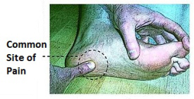
Causes
When a nerve is pinched between bones, the result is swelling of the nerve. It is this swelling which is referred to as a Neuroma. When the condition occurs in the foot, it is known as a Morton?s Neuroma. Morton?s Neuroma is technically not a tumor. Rather, it is a thickening of the tissue that surrounds the digital nerves leading to the toes. These nerves allow for physical sensation on the skin of the toes. The region of inflammation is found where the digital nerve passes under the ligament connecting the toe bones (metatarsals) in the forefoot. Morton?s Neuroma commonly develops between the third and fourth toes, generally as a result of ongoing irritation, trauma or excessive pressure. In some cases, the second and third toes are involved. Morton?s Neuroma is confined to one foot in most cases, though it can occur in both, particularly in athletes such as runners.
Symptoms
People with Morton's neuroma usually complain of pain that can start in the ball of the foot and shoot into the affected toes. However, some people just have toe pain. There may also be burning and tingling of the toes. The symptoms are usually felt up the sides of the space between two toes. For example, if the nerve between the third and fourth long bones (metatarsals) of the right foot is affected, the symptoms will usually be felt up the right-hand side of the fourth toe and up the left-hand side of the third toe. Some people describe the pain that they feel as being like walking on a stone or a marble. Symptoms can be made worse if you wear high-heeled shoes. The pain is relieved by taking your shoe off, resting your foot and massaging the area. You may also experience some numbness between the affected toes. Your affected toes may also appear to be spread apart, which doctors refer to as the 'V sign'. The symptoms can vary and may come and go over a number of years. For example, some people may experience two attacks of pain in a week and then nothing for a year. Others may have regular and persistent (chronic) pain.
Diagnosis
The doctor will ask about your symptoms and medical history. A physical exam will be done. Initial diagnosis of Morton's neuroma is based on your description of the type and location of pain and discomfort in the foot. The diagnosis will be confirmed by a physical exam of the foot, including checking for mechanical abnormalities in the foot, squeezing the side of the foot, which will usually cause pain when Morton's neuroma is present. Examination of your shoes to check for excess wear in parts of the shoe, check to see whether the shoes are too tight. Imaging tests evaluate the foot and surrounding structures. This may be done with X-ray, MRI scan, Ultrasound. Injections of local anesthetic can also be used for diagnosis.
Non Surgical Treatment
Conservative treatment for Morton?s neuroma involves footwear that allows your forefoot to spread. High-heeled shoes cause neuromas by squeezing and stretching your involved intermetatarsal nerve across the ball of your foot and should be avoided as often as possible. A shoe that possesses any toe spring will also place more stress on your foot nerves and increase your likelihood of developing a neuroma. Test shoes before you buy them to see if they are appropriate for your feet. Select shoes that have a removable liner or insole, and stand on the liner, noting the position of your foot. If your foot is wider than your liner, that shoe will irritate your neuroma by squeezing your metatarsal bones together.
Surgical Treatment
If pain persists with conservative care, surgery may be an appropriate option. The common digitial nerve is cut and the Mortons neuroma removed. This will result is numbness along the inside of the toes affected, and there is a small chance the end of the nerve will form a Stump Neuroma. Approximately 75% of people receive symptom resolution for Mortons Neuroma with conservative care.
A Morton?s Neuroma is actually incorrectly termed, with the name suggesting it is a tumour or growth. Rather than a true neuroma it is actually what is called a perineural fibrosis, which means that over time the sheath surrounding the nerve becomes irritated, inflamed, and forms a thickened scar tissue.Causes
When a nerve is pinched between bones, the result is swelling of the nerve. It is this swelling which is referred to as a Neuroma. When the condition occurs in the foot, it is known as a Morton?s Neuroma. Morton?s Neuroma is technically not a tumor. Rather, it is a thickening of the tissue that surrounds the digital nerves leading to the toes. These nerves allow for physical sensation on the skin of the toes. The region of inflammation is found where the digital nerve passes under the ligament connecting the toe bones (metatarsals) in the forefoot. Morton?s Neuroma commonly develops between the third and fourth toes, generally as a result of ongoing irritation, trauma or excessive pressure. In some cases, the second and third toes are involved. Morton?s Neuroma is confined to one foot in most cases, though it can occur in both, particularly in athletes such as runners.
Symptoms
People with Morton's neuroma usually complain of pain that can start in the ball of the foot and shoot into the affected toes. However, some people just have toe pain. There may also be burning and tingling of the toes. The symptoms are usually felt up the sides of the space between two toes. For example, if the nerve between the third and fourth long bones (metatarsals) of the right foot is affected, the symptoms will usually be felt up the right-hand side of the fourth toe and up the left-hand side of the third toe. Some people describe the pain that they feel as being like walking on a stone or a marble. Symptoms can be made worse if you wear high-heeled shoes. The pain is relieved by taking your shoe off, resting your foot and massaging the area. You may also experience some numbness between the affected toes. Your affected toes may also appear to be spread apart, which doctors refer to as the 'V sign'. The symptoms can vary and may come and go over a number of years. For example, some people may experience two attacks of pain in a week and then nothing for a year. Others may have regular and persistent (chronic) pain.
Diagnosis
The doctor will ask about your symptoms and medical history. A physical exam will be done. Initial diagnosis of Morton's neuroma is based on your description of the type and location of pain and discomfort in the foot. The diagnosis will be confirmed by a physical exam of the foot, including checking for mechanical abnormalities in the foot, squeezing the side of the foot, which will usually cause pain when Morton's neuroma is present. Examination of your shoes to check for excess wear in parts of the shoe, check to see whether the shoes are too tight. Imaging tests evaluate the foot and surrounding structures. This may be done with X-ray, MRI scan, Ultrasound. Injections of local anesthetic can also be used for diagnosis.
Non Surgical Treatment
Conservative treatment for Morton?s neuroma involves footwear that allows your forefoot to spread. High-heeled shoes cause neuromas by squeezing and stretching your involved intermetatarsal nerve across the ball of your foot and should be avoided as often as possible. A shoe that possesses any toe spring will also place more stress on your foot nerves and increase your likelihood of developing a neuroma. Test shoes before you buy them to see if they are appropriate for your feet. Select shoes that have a removable liner or insole, and stand on the liner, noting the position of your foot. If your foot is wider than your liner, that shoe will irritate your neuroma by squeezing your metatarsal bones together.
Surgical Treatment
If pain persists with conservative care, surgery may be an appropriate option. The common digitial nerve is cut and the Mortons neuroma removed. This will result is numbness along the inside of the toes affected, and there is a small chance the end of the nerve will form a Stump Neuroma. Approximately 75% of people receive symptom resolution for Mortons Neuroma with conservative care.
Treating Leg Length Imbalances With Shoe Lifts
There are actually not one but two different kinds of leg length discrepancies, congenital and acquired. Congenital means you are born with it. One leg is anatomically shorter in comparison to the other. As a result of developmental stages of aging, the brain picks up on the stride pattern and recognizes some difference. The body usually adapts by dipping one shoulder to the "short" side. A difference of less than a quarter inch isn't very excessive, does not need Shoe Lifts to compensate and in most cases does not have a serious effect over a lifetime.

Leg length inequality goes typically undiagnosed on a daily basis, however this issue is simply solved, and can eliminate many cases of lumbar pain.
Treatment for leg length inequality usually consists of Shoe Lifts . These are very inexpensive, regularly priced at below twenty dollars, compared to a custom orthotic of $200 plus. Differences over a quarter inch can take their toll on the spine and should probably be compensated for with a heel lift. In some cases, the shortage can be so extreme that it requires a full lift to both the heel and sole of the shoe.
Mid back pain is easily the most common health problem affecting men and women today. Around 80 million people are afflicted by back pain at some point in their life. It's a problem that costs employers millions every year due to time lost and productivity. Fresh and better treatment methods are continually sought after in the hope of minimizing the economical influence this condition causes.

People from all corners of the world suffer from foot ache due to leg length discrepancy. In these types of situations Shoe Lifts are usually of beneficial. The lifts are capable of alleviating any pain and discomfort in the feet. Shoe Lifts are recommended by many professional orthopaedic doctors.
To be able to support the human body in a well balanced fashion, feet have a very important part to play. In spite of that, it's often the most overlooked region in the human body. Some people have flat-feet which means there may be unequal force placed on the feet. This causes other areas of the body like knees, ankles and backs to be affected too. Shoe Lifts ensure that the right posture and balance are restored.
Leg length inequality goes typically undiagnosed on a daily basis, however this issue is simply solved, and can eliminate many cases of lumbar pain.
Treatment for leg length inequality usually consists of Shoe Lifts . These are very inexpensive, regularly priced at below twenty dollars, compared to a custom orthotic of $200 plus. Differences over a quarter inch can take their toll on the spine and should probably be compensated for with a heel lift. In some cases, the shortage can be so extreme that it requires a full lift to both the heel and sole of the shoe.
Mid back pain is easily the most common health problem affecting men and women today. Around 80 million people are afflicted by back pain at some point in their life. It's a problem that costs employers millions every year due to time lost and productivity. Fresh and better treatment methods are continually sought after in the hope of minimizing the economical influence this condition causes.
People from all corners of the world suffer from foot ache due to leg length discrepancy. In these types of situations Shoe Lifts are usually of beneficial. The lifts are capable of alleviating any pain and discomfort in the feet. Shoe Lifts are recommended by many professional orthopaedic doctors.
To be able to support the human body in a well balanced fashion, feet have a very important part to play. In spite of that, it's often the most overlooked region in the human body. Some people have flat-feet which means there may be unequal force placed on the feet. This causes other areas of the body like knees, ankles and backs to be affected too. Shoe Lifts ensure that the right posture and balance are restored.
Are Shoe Lifts The Ideal Solution To Leg Length Imbalances
There are two different kinds of leg length discrepancies, congenital and acquired. Congenital indicates you are born with it. One leg is structurally shorter compared to the other. Through developmental phases of aging, the brain senses the gait pattern and recognizes some variance. The entire body usually adapts by dipping one shoulder to the "short" side. A difference of less than a quarter inch isn't really excessive, demand Shoe Lifts to compensate and usually won't have a serious effect over a lifetime.

Leg length inequality goes typically undiagnosed on a daily basis, yet this condition is very easily solved, and can eradicate numerous cases of back discomfort.
Treatment for leg length inequality commonly involves Shoe Lifts. These are typically cost-effective, usually priced at less than twenty dollars, in comparison to a custom orthotic of $200 plus. When the amount of leg length inequality begins to exceed half an inch, a whole sole lift is generally the better choice than a heel lift. This prevents the foot from being unnecessarily stressed in an abnormal position.
Upper back pain is the most prevalent condition impacting men and women today. Over 80 million men and women are affected by back pain at some stage in their life. It is a problem that costs employers vast amounts of money each year as a result of time lost and productivity. New and more effective treatment solutions are always sought after in the hope of reducing the economical influence this condition causes.

Men and women from all corners of the world suffer the pain of foot ache due to leg length discrepancy. In these cases Shoe Lifts can be of worthwhile. The lifts are capable of alleviating any discomfort and pain in the feet. Shoe Lifts are recommended by many experienced orthopaedic practitioners".
In order to support the human body in a healthy and balanced fashion, the feet have a very important task to play. In spite of that, it can be the most neglected zone of the human body. Many people have flat-feet meaning there may be unequal force placed on the feet. This will cause other parts of the body including knees, ankles and backs to be impacted too. Shoe Lifts ensure that proper posture and balance are restored.
Leg length inequality goes typically undiagnosed on a daily basis, yet this condition is very easily solved, and can eradicate numerous cases of back discomfort.
Treatment for leg length inequality commonly involves Shoe Lifts. These are typically cost-effective, usually priced at less than twenty dollars, in comparison to a custom orthotic of $200 plus. When the amount of leg length inequality begins to exceed half an inch, a whole sole lift is generally the better choice than a heel lift. This prevents the foot from being unnecessarily stressed in an abnormal position.
Upper back pain is the most prevalent condition impacting men and women today. Over 80 million men and women are affected by back pain at some stage in their life. It is a problem that costs employers vast amounts of money each year as a result of time lost and productivity. New and more effective treatment solutions are always sought after in the hope of reducing the economical influence this condition causes.
Men and women from all corners of the world suffer the pain of foot ache due to leg length discrepancy. In these cases Shoe Lifts can be of worthwhile. The lifts are capable of alleviating any discomfort and pain in the feet. Shoe Lifts are recommended by many experienced orthopaedic practitioners".
In order to support the human body in a healthy and balanced fashion, the feet have a very important task to play. In spite of that, it can be the most neglected zone of the human body. Many people have flat-feet meaning there may be unequal force placed on the feet. This will cause other parts of the body including knees, ankles and backs to be impacted too. Shoe Lifts ensure that proper posture and balance are restored.
Have I Got Posterior Calcaneal Spur

Overview
The heel spur (or calcaneal spur) is a nail-like growth of calcium around the ligaments and tendons of the foot where they attach to the heel bone. The spur grows from the bone and into the flesh of the foot. A heel spur results from an anatomical change of the calcaneus (heel bone). This involves the area of the heel and occasionally, another disability, such as arthritis. The heel bone forms one end of the two longitudinal arches of the foot. These arches are held together by ligaments and are activated by the muscles of the foot (some of which are attached beneath the arches and run from the front to the back of the foot). These muscles and ligaments, like the other supporting tissues of the body, are attached in two places. Many are attached at the heel bone. The body reacts to the stress at the heel bone by calcifying the soft tissue attachments and creating a spur.
Causes
The main cause of heel spur is calcium deposit under the heel bone. Building of calcium deposits can take place over several months. Heel spurs happens because of stress on the foot ligaments and muscles and continuous tearing of the membrane covering the heel bone. It also happens due to continuous stretching the plantar fascia. Heel spurs are mostly seen in case of athletes who has to do lots of jumping and running. The risk factors that may lead to heel spurs include aormalities in walking which place too much stress on the heel bone, nerves in the heel and ligaments. Poorly fitted shoes without the right arch support. Jogging and running on hard surfaces. Excess weight. Older age. Diabetes. Standing for a longer duration.

Symptoms
Pain and discomfort associated with heel spurs does not occur from the spur itself. The bone growth itself has no feeling. However, as you move, this growth digs into sensitive nerves and tissue along the heel of the foot, resulting in severe pain. Pain can also be generated when pushing off with the toes while walking. Swelling along the heel is also common.
Diagnosis
Your doctor will review your medical history and examine your foot. X-rays are used to identify the location and size of the heel spur.
Non Surgical Treatment
In extreme cases, a doctor may recommend surgery for the removal of heel spurs. Fortunately, this is the exception rather than the rule. Most cases can be resolved with a combination of icing, rest, foot stretches and supporting the foot with an orthodic shoe insert specifically designed for this condition. We recommend that you continue on to our article on Heel Spur Treatment to discover the best, speediest and most affordable methods of resolving this ailment without invasive medical procedures.
Surgical Treatment
In some cases, heel spurs are removed by surgery after an X-ray. While the surgery is typically effective, it?s a timely and expensive procedure. Even after surgery, heel spurs can re-form if the patient continues the lifestyle that led to the problem. These reasons are why most people who develop painful heel spurs begin looking for natural remedies for joint and bone pain. Surgery isn?t required to cure a heel spur. In fact, more than 90 percent of people get better with nonsurgical treatments. If nonsurgical methods fail to treat symptoms of heel spurs after 12 months, surgery may be necessary to alleviate pain and restore mobility.
Prevention
There are heel spur prevention methods available in order to prevent the formation of a heel spur. First, proper footwear is imperative. Old shoes or those that do not fit properly fail to absorb pressure and provide the necessary support. Shoes should provide ample cushioning through the heel and the ball of the foot, while also supporting the arch. Wearing an orthotic shoe insert is one of the best ways to stretch the plantar fascia and prevent conditions such as heel spurs. Stretching the foot and calf is also helpful in preventing damage. Athletes in particular should make sure to stretch prior to any physical activity. Stretching helps prevent heel spurs by making tissue stronger as well as more flexible. In addition, easing into a new or increasingly difficult routine should be done to help avoid strain on the heel and surrounding tissue.